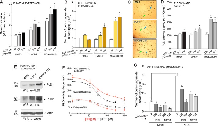
FIGURE 1.
PLD is important for breast cancer cell invasion. A, PLD gene expression was measured by quantitative RT-PCR in three cell lines after stimulation with 30 nm EGF for 0, 3, and 6 h. B, cell invasion was measured in HMEC, MCF-7, and MDA-MB-231 cells for 3 and 6 h in response to 30 nm EGF as chemoattractant. Results are expressed in terms of the mean total number of cells invading the Matrigel insert ±S.E. for each time point. Cell invasion assays were performed in duplicate, and ∼six fields were counted per insert. C, actual photographs (20× magnification) of representative fields for invaded cells (after 6 h) in Matrigel. Green arrows point at round, empty circles that are the membrane pores. Blue arrows point at irregularly shaped, black spots, which are the stained cells that had invaded the Matrigel. D, enzymatic activity of endogenous PLD was measured in HMEC, MCF-7, and MDA-MB-231 cell lines. Results are expressed in terms of the mean % of total PLD activity compared with the mock control samples ±S.E. Duplicate sample were assayed with n = 3 total assays performed. E, endogenous protein expression of PLD1 and PLD2 was detected by Western blot (W.B.) analysis. β-Actin was used as a readout for equal protein loading control. F, both endogenous and overexpressed PLD enzymatic activity was measured in the presence of increasing amounts of PLD inhibitor, FIPI or NFOT. G, cell invasion was measured in MDA-MB-231 cells and overexpression of PLD2 and incubation with PLD inhibitor, FIPI or NFOT.

