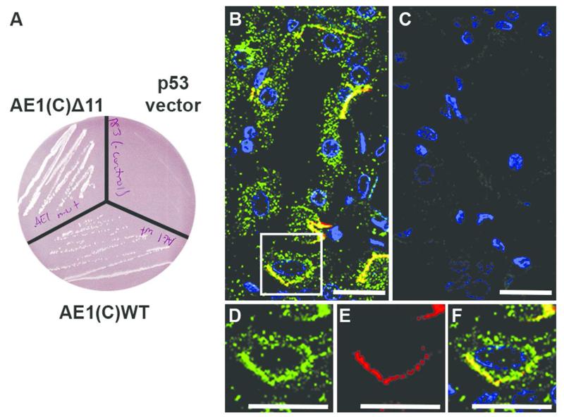
Figure 1. Confirmation of PRDX6 as binding candidate of kAE1and co-staining of intercalated cell.
(A) Yeast mating test of both AE1(C)WT and AE1(C)Δ11 with PRDX6 in yeast cells shows protein potential interaction indicated by the blue colonies on selection plates with no cell growth of the negative control (p53 empty vector). (B-F) Immunofluorescence in human renal collecting duct. PRDX6 (green) and AE1 (red) co-localize in a number of collecting duct cells (B) with absence of staining when primary antisera were omitted (C). An enlarged image of an intercalated cell shows widespread PRDX6 expression (D) and basolateral AE1 (Bric 170) expression (E). Merged panel (F) indicates co-localization of PRDX6 and AE1 at the basolateral surface. (Scale bars = 20 μm)

