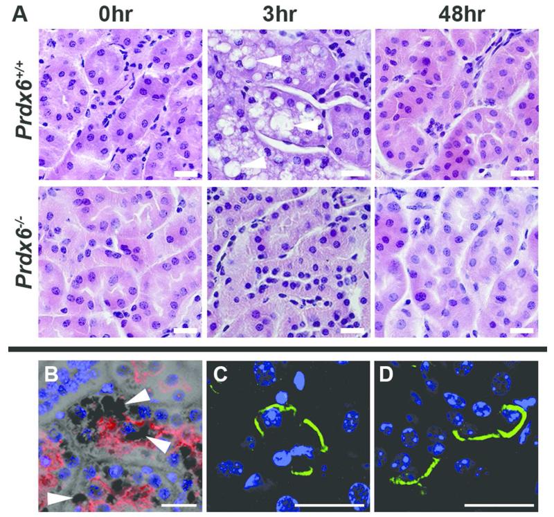
Figure 4. Cellular changes in acidified adult Prdx6+/+ and Prdx6−/− mouse kidney.
(A) H+E stained mouse renal cortex of Prdx6+/+ and Prdx6−/− at baseline, 3 hours and 48 hours after acid gavage showing tubular vacuolation in Prdx6+/+ animals (white arrowheads) which later resolved. This vacuolation was not evident in the cortex of Prdx6−/− animals after the same treatment. (B) Megalin staining (red) confirmed the proximal tubular location of the vacuoles (white arrowheads). AE1’s basolateral location (green) was similarly maintained in Prdx6+/+ (C) and Prdx6−/− (D) animals. (Scale bar = 20 μm)

