Figure 6.
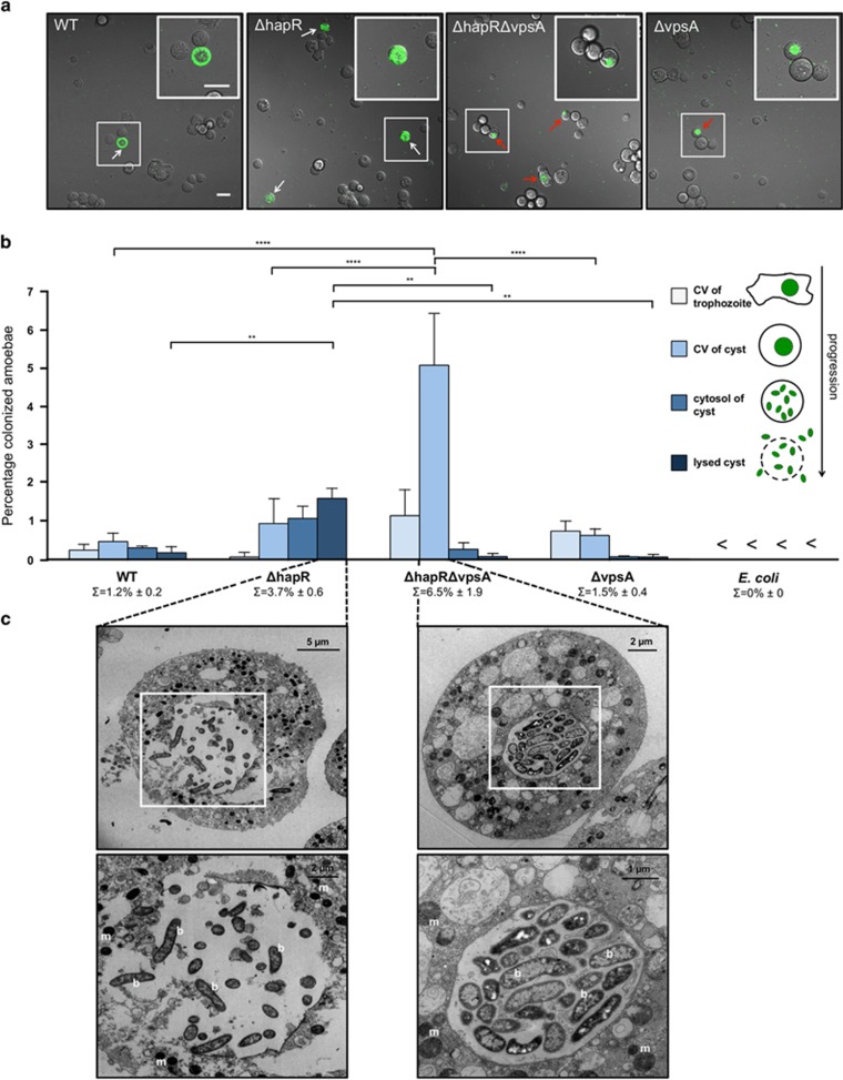
The VPS contributes to CV lysis. (a) V. cholerae strains lacking VPS are severely impaired in amoebal CV lysis. Confocal imaging of amoebal cysts formed in the presence of V. cholerae WT strain (WT) or mutant V. cholerae strains lacking hapR (ΔhapR), vpsA (ΔvpsA) or hapR and vpsA (ΔhapRΔvpsA). White arrows depict V. cholerae within the cytosol of the cysts after CV lysis and red arrows indicate colonized but intact CVs. Shown are merged images of the transmitted light and the GFP channels taken at 20 h p.p.c. The insets show magnifications of the boxed regions. Scale bars: 20 μm. (b) Quantification of specific events during amoebal colonization. A. castellanii cells harboring V. cholerae inside the CV of the trophozoite, the CV of the cyst and within the cytosol of the cyst before and after cyst lysis were counted. Values represent averages from three independent experiments (4200 total amoebae counted for each strain). <, below the detection limit of 0.02%. Statistically significant differences are indicated (****P<0.0001; **P<0.01). ∑ indicates the total percentage of colonized CVs per V. cholerae strain (±s.d.). (c) Representative transmission electron micrographs showing colonized compartments of A. castellanii. V. cholerae strains ΔhapR and ΔhapRΔvpsA were cocultured with A. castellanii for 20 h before infected cysts were visualized by electron microscopy. The lower images are magnifications of the boxed regions in the upper images. b, bacteria; m, mitochondria. V. cholerae strains used for all panels were producing fluorescent proteins.

