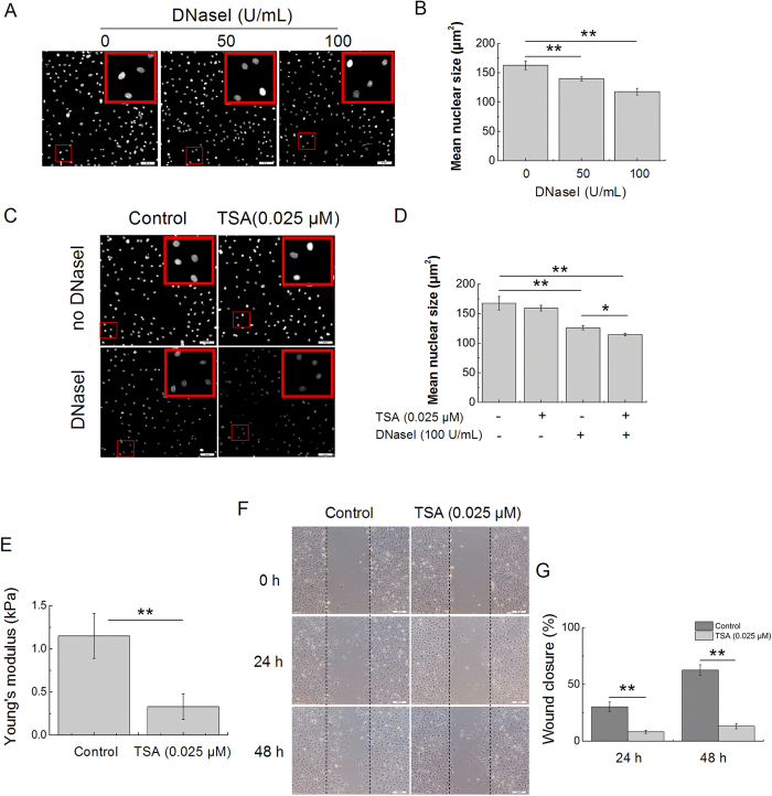
Figure 4. Chromatin decondensation reduces nuclear stiffness and cell migration.
(A) In situ DNaseI-sensitivity assay. Photomicrographs of DAPI-stained nuclei after 20 minutes of digestion with the different concentrations of DNaseI. Bars = 100 μm. (B) Quantitative analysis of the mean size of nuclei after DNaseI as shown in (A). n = 3 in each group. (C) Validation of the in situ DNaseI-sensitivity assay: decondensation of chromatin enhances the rate of digestion. Photomicrographs show that the nuclear sizes of the TSA-treated cells are significantly decreased when compared to those of the control cells. Bars = 100 μm. (D) Quantitative analysis of the mean size of nuclei after DNaseI as shown in (C). n = 3 in each group. (E) The nuclei of tenocytes exposed to TSA (0.025 μM) are less stiff than those of the control cells (non-TSA-treated). n = 15 in each group. (F) Images of the migration of tenocytes treated with TSA, as evaluated through a scratch wound assay. Bars = 200 μm. (G) Percentages of wound closure obtained for tenocytes treated with TSA. n = 3 in each group. The data are expressed as the means ± SD; *p < 0.05 and **p < 0.01.

