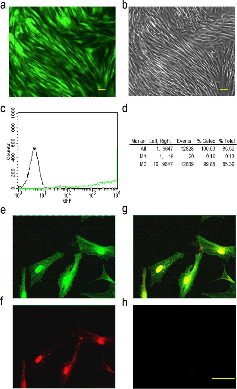
Figure 2. Infection of Ad-Runx2 (co-expression with EGFP) and expression of Runx2 in ADSCs.
(a,b) Runx2 expression (green) in Ad-Runx2-infected ADSCs at 48 h post-infection was examined by fluorescence microscopy (a) and control cell morphology by light microscopy (b). (c,d) Ad-Runx2 transduction efficiency was measured by flow cytometry at 48 h post-infection (c) and using non-transduced ADSCs as controls for autofluorescence (d). (e–h) Immunofluorescence staining showed nuclear expression of Runx2 in Ad-Runx2-ADSCs at 48 h post-infection. EGFP (green) is expressed in the nucleus and cytoplasm (e), and Runx2 expression assayed by immuno-histology (red) is confined to the nucleus in infected cells (f). A merged image of (e,f) is shown in (g). Non-transduced cells were used as a negative control (h). The experiments were performed three times and representative images are shown. The scale bar is 20 μm.

