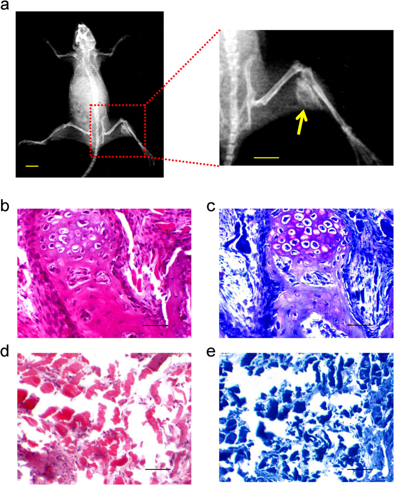
Figure 4. Ad-Runx2-infected ADSCs promote ectopic bone formation in vivo.
(a) Bone formation was detected by X-rays in the muscle of right lower limb, 8 weeks after intramuscular injection of Ad-Runx2-modified ADSCs into the right lower limb of nude mice, but not in the left control limb injected with ADSCs infected with Ad-EGFP (scale bar is 10 mm). (b–e) Histological analyses of in vivo osteogenesis by Ad-Runx2-modified ADSCs. Note that significant amounts of cartilage and bone formation were observed with hematoxylin & eosin (b) and toluidine blue staining (c) in the Ad-Runx2-ADSCs group. The Ad-EGFP-ADSCs group contained muscle and fibrous tissue, with no evidence of bone formation (d,e). Representative images from six replications are shown.

