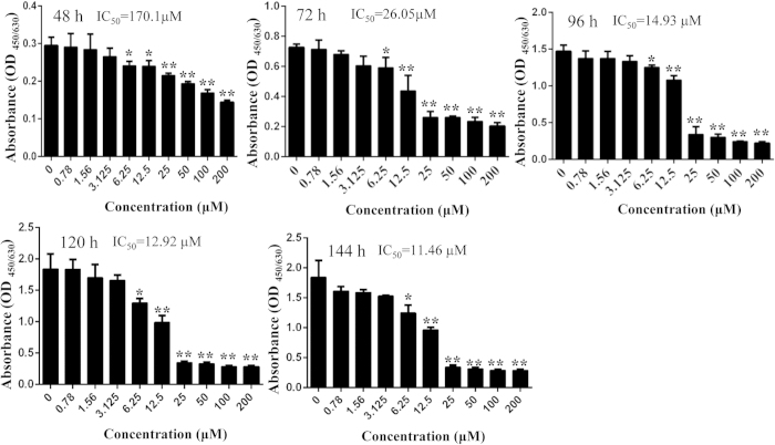
Figure 2. DHA inhibited osteoclastogenesis without toxicity to BMMs.
BMMs were treated with indicated concentrations of DHA, 30 ng/mL M-CSF, and 50 ng/mL RANKL for 48 h, 72 h, 96 h, 120 h and 144 h, respectively, and cell viability was then measured using CCK-8 assay. The IC50 for DHA in BMMs at 48 h, 72 h, 96 h, 120 h and 144 h were also calculated. (*p < 0.05, **p < 0.01).

