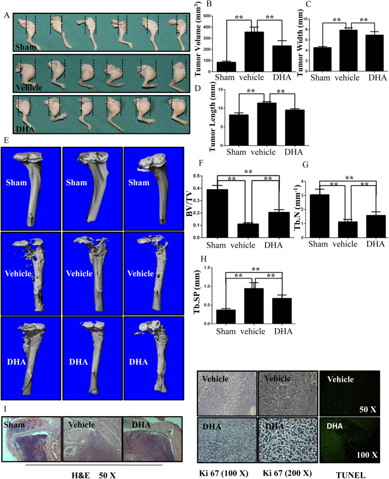
Figure 8. DHA inhibits breast cancer bone metastasis and osteolysis in vivo.
(A) MDA-MB-231 cells were injected directly into the tibiae plateau. After 28 days, the tissue volume (B), tissue width (C) and tissue length (D) were measured in the vehicle group. (E) Representative micro-CT and 3-dimensional reconstructed images from each group are shown. (F) The BV/TV of each sample was measured. (G) Trabecular number (Tb.N) of each sample was measured. (H) Trabecular separation (Tb.Sp) of each sample was measured. (I) The tumor volume sections were fixed, decalcified, dehydrated, and sectioned. Sections were stained with H&E, Ki67 and TUNEL. (*p < 0.05, **p < 0.01).

