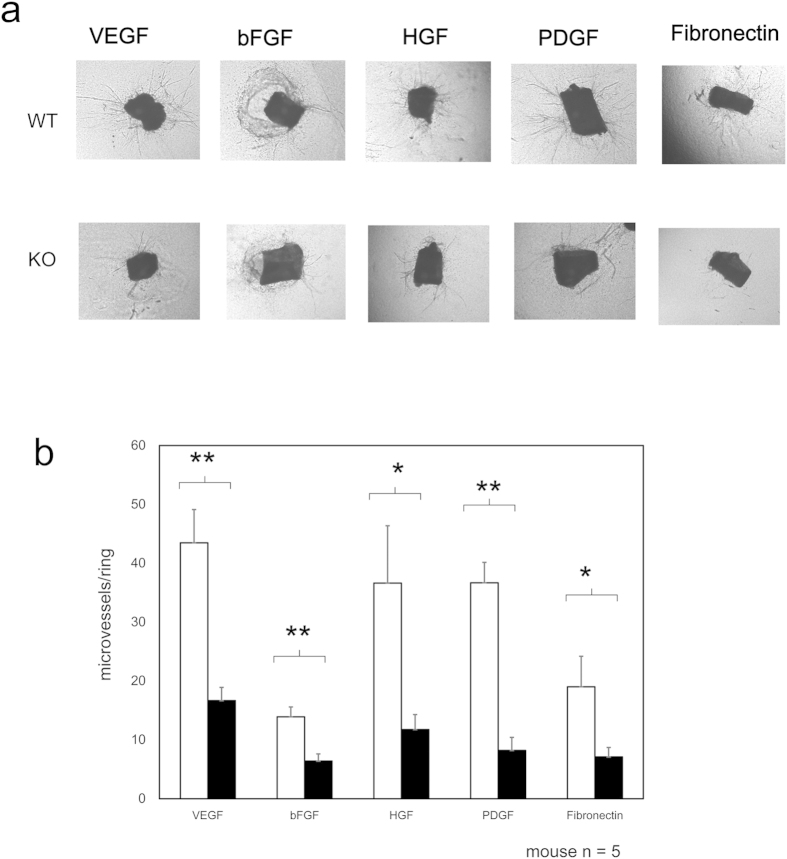
Figure 3. Influence of PKN3 KO in the regulation of ex vivo angiogenesis.
(a) Abdominal aortic ring segments from WT or PKN3 KO mice embedded in matrigel (for PDGF) or collagen (for VEGF, bFGF, HGF, and Fibronectin). Aortic ring segments were incubated with each growth factor indicated for 6 days. Panel shows representative photomicrographs of microvascular sprouting in each condition after 6 days in culture. (b) Effect on the sprouting vessels from ex vivo aortic rings. Bars represent mean of 15 independent experiments ± SEM. (mouse number of each genotype is 5). * and ** indicate P < 0.05 and P < 0.01, respectively.

