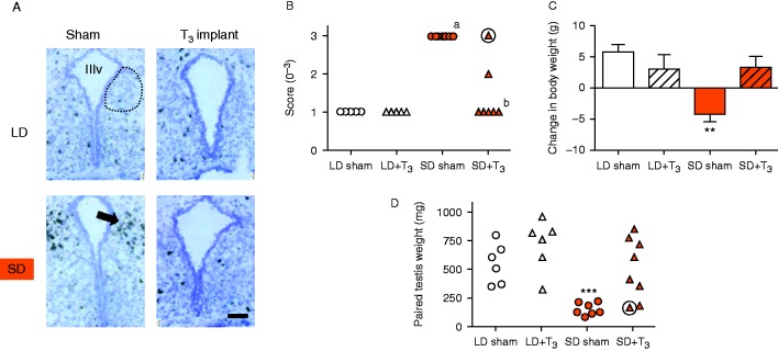
Figure 4.
Intra-hypothalamic T3 administration reduces VGF mRNA expression in the SD Siberian hamster. (A) representative photomicrographs of coronal sections through the dmpARC counterstained with cresyl violet, VGF hybridization is revealed by dark silver grains in the overlying emulsion in Siberian hamsters exposed to LD or SD receiving intra-hypothalamic sham or T3 implants for 8 weeks. Dotted line indicates approximate boundaries of the dmpARC, arrow indicates induces expression in a SD sham hamster, scale bar=100 μm. (B) analysis of VGF mRNA abundance, scores for individual animals are depicted; aP<0.001 vs LD sham group, bP<0.05 vs SD sham group. (C) overall change in body weight, values are mean±s.e.m., **P<0.01 vs LD-sham group. (D) individual paired testis weights at the end of the study, ***P<0.001 vs LD-sham group. Weekly mean body weight data and group mean testis weight data have been published previously (Barrett et al. 2007). Circled values (panels (B) and (D)) are data from the same individual.

 This work is licensed under a