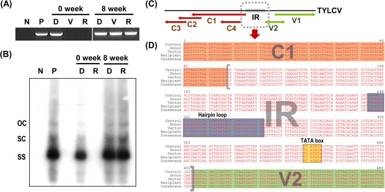
Figure 5. PCR analysis confirming whitefly-mediated transmission of seed-borne TYLCV from infected to healthy tomato plants.
(A) PCR analysis using a TYLCV-specific primer set and (B) Southern hybridization using a TYLCV-specific probe at 0 and 8 weeks after whitefly release in tents. Lane N, negative control; lane P, positive control with TYLCV-infected tomato genomic DNA; lane D, donor plant; lane V, vector (whitefly); and lane R, recipient plant genomic DNA. OC, open-circular double-stranded DNA; SC, supercoiled double-stranded DNA; SS, single-stranded DNA. (C) Linearized diagram of TYLCV DNA. Red and green arrows indicate coding sequences and the gray dotted box indicates the region used for sequence analysis containing the intergenic region of TYLCV. (D) Multiple sequence alignments of the TYLCV partial genome from donor and recipient plants and a vector with the control sequence (JN680149).

