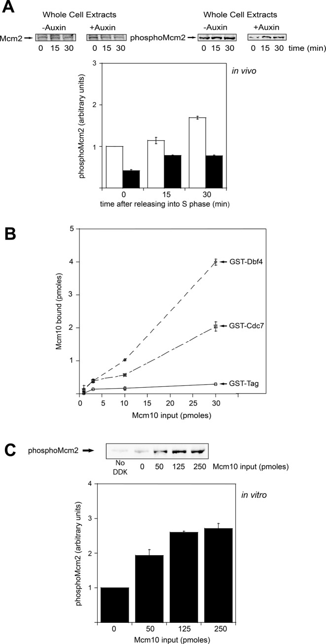
Figure 3.
Mcm10 interacts with DDK and stimulates Mcm2 phosphorylation by DDK. (A) mcm10-1-aid cells were grown as described in Materials and Methods. (A, left panel) Whole cell extracts were analyzed by western blot for expression of Mcm2. (A, right panel) Whole cell extracts were analyzed by western blot for expression of phospho-Mcm2. Results from similar experiments were quantified, averaged and plotted. (B) 30 pmol of GST-Dbf4, GST-Cdc7 or GST tag was incubated with increasing concentrations of radiolabeled PKA-Mcm10 at 30°C for 10 min in a GST pulldown assay. Results from similar experiments were quantified, averaged and plotted. (C) 5 μg of Mcm2 was incubated with 50 ng of DDK and varying amounts of Mcm10 in a volume of 25 μl at 30°C for 1 h. The reactions were then analyzed by western blot for expression of phospho-Mcm2. Results from similar experiments were quantified, averaged and plotted. Graphs from (A), (B) and (C) represent mean values from two independent experiments and error bars indicate the standard deviation of the mean.

