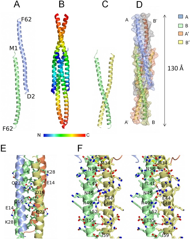
Figure 2.
Structure of the YabA-NTD tetramer. (A) Ribbon representation of the two chains of YabA1–62 in the asymmetric unit A (ice blue) and B (light green) with the N- and C-terminal residues labelled. (B and D) The YabA1–62 tetramer. Each chain is shown as a ribbon either colored ramped as shown in the key (B) or colored by chain (D) with an accompanying surface rendering. (C) Ribbon representation of molecules A’ and B illustrating the tweezer arrangement of these chains. (E) The four helix bundle region. The chains are displayed as ribbons colored by chain A (ice blue) B (light green) A’ (lemon) and B’ (coral). The Cα and side chains of apolar residues in the interior are colored by residue type, light green for Val and Ile and lawn green for Leu to emphasize the leucine zipper. Polar interactions between the chains are shown as dashed lines and residues forming the prominent interchain interactions referred to in the text are labeled. (F) Stereo image of the coiled coil region of chains B and A’. The Cα and the side chains of residues from the two chains are displayed with the carbon atoms colored according to the chain and with nitrogens in blue, oxygens in red and sulphurs in yellow. Polar interactions between the chains are shown as dashed lines.

