
Figure 5.
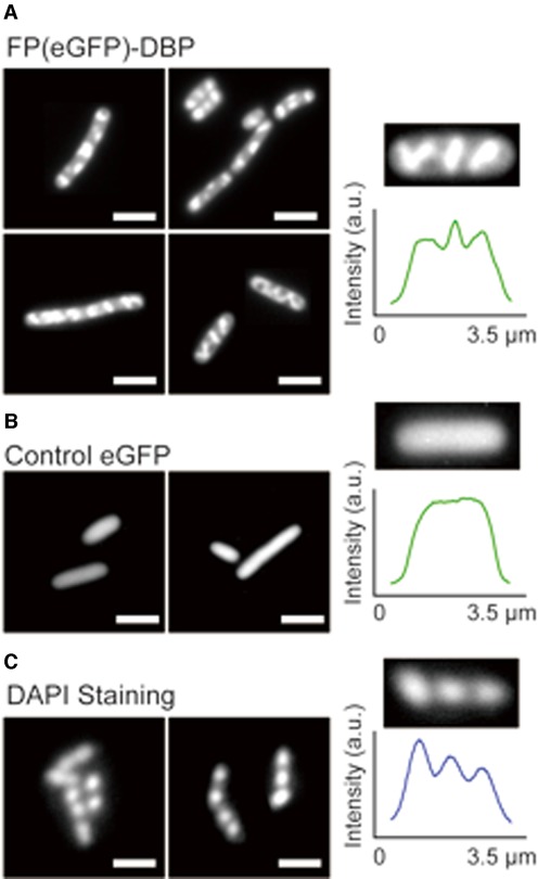
Live E. coli cells are specifically stained by endogenously expressed FP-DBP. (A) E. coli cells (BL21) expressing FP(eGFP)-DBP show discrete patterns within bacterial cells. Images show localized fluorescence features within E. coli cells; a fluorescence intensity profile across one cell details this pattern. (B) Control: E. coli cells expressing eGFP, but without the KWKWKKA peptide, and its featureless fluorescence intensity profile. (C) E. coli cells stained with DAPI, showing similar patterns of nucleoids. Scale bars = 5 μm.