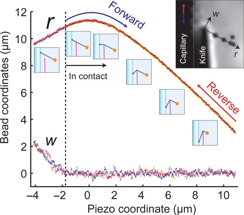
Figure 3.

Bead motion in the DNA pulley. The piezo coordinate p is positive for downward motion of the capillary. For P < −2 mm, the DNA was out of contact with the blade. Initially the w- and r-coordinates varied with P according to their projection along the direction of piezo motion. For −2 mm < P < 0 mm, r increased as the DNA-capillary junction approached the blade. Thereafter the DNA-capillary junction receded from the blade and the bead was pulled toward the blade. Blue and red points represent measurements of bead location during a forward and reverse scan, respectively. Yellow line represents a fit to Equation (1).
