
Figure 2.
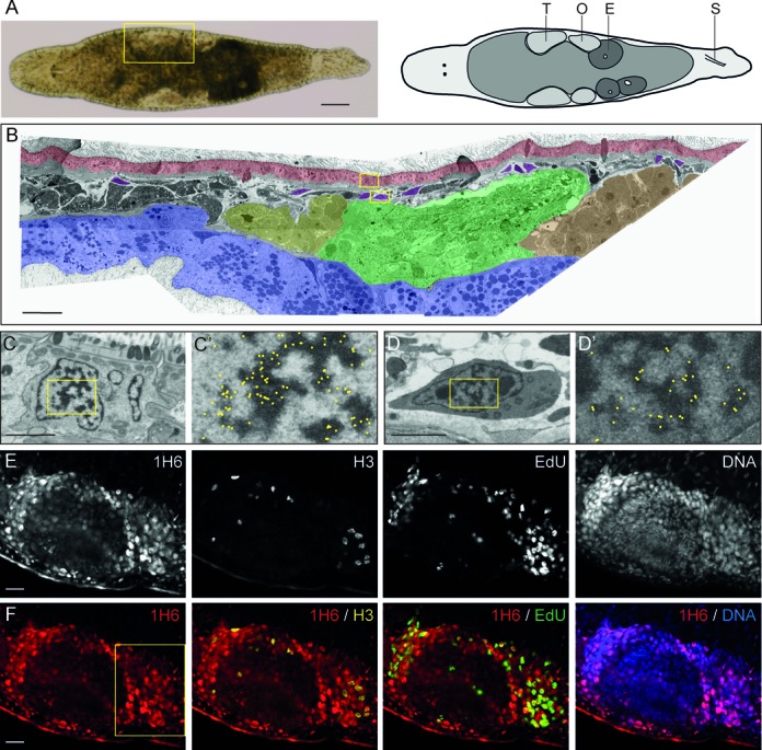
Weak 1H6 staining of proliferating cells in adult Macrostomum. (A) Bright-field image and schematic overview of Macrostomum lignano. T: Testis, O: Ovary, E: Egg and S: Stylet. (B) False-colored overview of 1H6 immuno-EM nanotomy data of Figure 2 available at http://www.nanotomy.org/. Red: Epidermis; Blue: Gut; Yellow: Testis Tip; Green: Testis; Orange: Ovary; Purple: somatic stem cells (neoblasts). (C–D’) EM images of an epidermal cell (C, C’) and a neoblast (D, D’) following labeling with 1H6 antibody. Boxed areas in B are shown in C and D. Boxed area in C and D are shown in C’ and D’. For clarity, the immunogold particles are marked manually with yellow dots in C’ and D’ (raw data available online). (E) Gray scale images of a section through the germline. stained with 1H6, EdU (S-phase cells), phospho-H3 (mitotic cells) and DNA. (F) Colored overlays of sections shown in E. Higher magnifications of the boxed area in F are shown in Supplementary Figure S3. Scale bars: A: 100 μm, B: 30 μm; C, D: 2 μm; E–F: 20 μm.