
Figure 6.
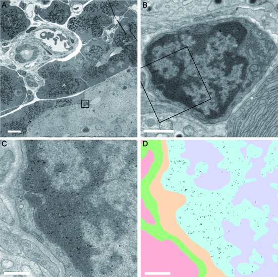
1H6 binds to heterochromatin in different cells of rat pancreas. (A) Overview of pancreatic tissue with alpha cells, beta cells, endothelial cells, epithelial cells and exocrine cells. The different cell types can be recognized as described (www.nanotomy.nl) (27). A high-resolution ‘nanotomy’ digital file of Figure 6, were gold-particles can be detected in a ‘Google-earth’ like analysis, is available online at http://www.nanotomy.org/). The boxed area in (A) is shown in (B). The boxed area in (B) is shown in (C) and (D). Annotation of the ultrastructure (C) is illustrated in (D) as follows: heterochromatin blue; euchromatin purple; cytoplasm yellow; extracellular space in green and an adjacent cell (cytoplasm) in pink. The data of panel (C) were processed to selectively visualize the gold particles (black). Bars: A: 10 μm; B: 1 μm; C,D: 0.5 μm.