Figure 1.

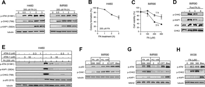
Activation of ATM pathway by FA. (A) ATM signaling in H460 and IMR90 cells treated with 200 μM FA for different time intervals. (B) Clonogenic survival of H460 cells treated with 200 μM FA for 1, 2 or 3 h. Means ± SD from two experiments each including 3 dishes/dose. (C) Cytotoxicity of 1-h and 3-h long FA treatments in IMR90 cells. Cell viability was measured at 72 h post-FA using the CellTiter-Glo assay. Means ± SD from two experiments with 3 dishes/dose. (D) ATM responses in IMR90 treated with 200 μM FA for 3 h and collected at the indicated recovery times. (E) Effects of ATM inhibitors on FA-induced protein phosphorylation (ATMi-1–KU55933, ATMi-2–KU60019). H460 cells were preincubated with inhibitors for 1 h and collected immediately after 3-h long FA treatments. (F) CHK1 and p53 phosphorylation in IMR90 cells treated for 3 h with FA, hydroxyurea (HU), camptothecin (CPT, 1 μM) or bleomycin (Bleo, 30 μg/ml). (G) ATM-related phosphorylation in IMR90 treated as in panel F. MSH2 was used as a loading control. (H) ATM signaling in WI38 normal cells treated with FA and bleomycin as IMR90 in panel F.
