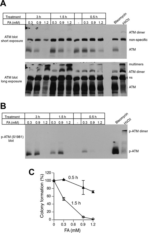
Figure 4.

Formation of inactive ATM dimers and multimers by high FA doses. H460 cells were treated with FA for 0.5–3 h or bleomycin (30 μg/ml) and H2O2 (8 mM) for 30 min. Proteins were denatured by the addition of 2% SDS without boiling and separated under nonreducing conditions at 4°C. (A) Western blot with anti-ATM antibodies. Short (top panel) and long (bottom panel) exposures of the same blot are shown. A non-specific band between ATM monomer and dimer was used as a loading control. (B) Western blot with anti-phospho-ATM antibodies. (C) Clonogenic survival of H460 cells treated with FA for 0.5 or 1.5 h. Data are Means ± SD, n=3.
