
Figure 3.
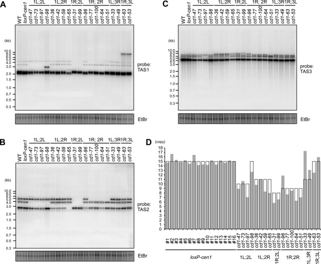
Alteration of subtelomere structures in the Δcen1 fusion survivors. (A–C) Southern blot analyses of genomic DNAs from the indicated telomere-fusion survivors. The DNAs were digested with NsiI and analysed by Southern blotting with the subtelomeric TAS1 (A), TAS2 (B) and TAS3 (C) probes. EtBr-stained images of the gels are shown as loading controls. The fusogenic end-dependent and survivor-specific band pattern alterations were observed by Southern blotting. In addition, the band intensity ratio was variable, even between survivors exhibiting the same band pattern. These findings suggest general subtelomere destabilization in the survivors. (D) The predicted and experimentally determined STE2 copy numbers in the indicated survivors and 16 independent loxP-cen1 clones of different ages (clones #1–16) are shown in the open and grey columns, respectively. Details of the STE2 copy number prediction are shown in Supplementary Figure S7A. For experimental STE2 copy number determination, the STE2 qPCR Ct value was normalized to that of act1+ in the same genome as a single-copy control, and was adjusted further by multiplying by a constant factor to ensure that the average STE2 copy number in the loxP-cen1 clones was equal to 15. The difference between the predicted and measured STE2 copy numbers was significantly larger in the survivors than the parental loxP-cen1 clones (P = 0.00017 by a Welch's two-tailed t-test; see also Supplementary Figure S9B).