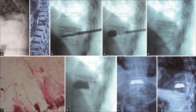
Figure 3.
71 year old female had a compression fracture of T11. (a) Standing lateral radiograph showing T11 fracture and local kyphosis (b) Magnetic resonance imaging showing the coexistence of both air and fluid in the T11 (c-d) Intraoperative fluoroscopy showing balloon inflated (e) Intraoperative fluoroscopy showing restoration of height of D11 (f) Histopathological report showing necrotic tissue. (g) Postoperative lateral fluoroscopy view showing well filled cement (h-i) Followup (18 months) x-rays anteroposterior and lateral views dorsal spine showing well filled cement and no dislodgement after surgery

