Figure 1.

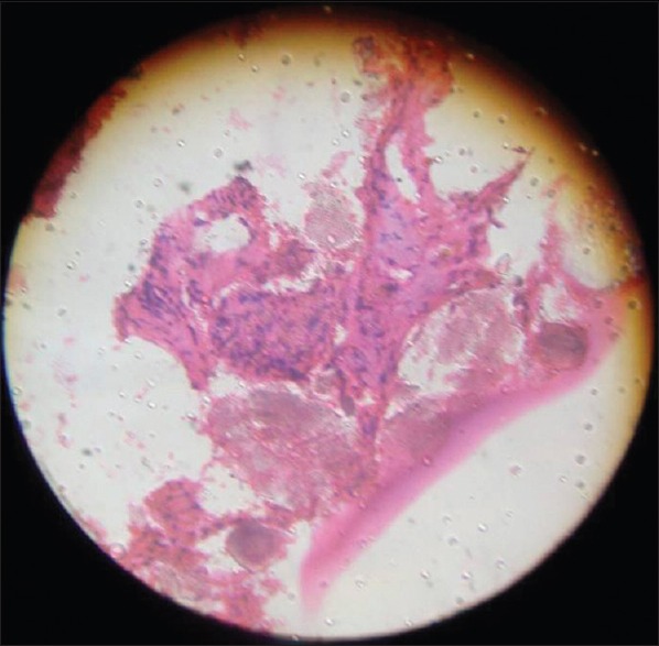
Histopatholgical picture showing new osteoid formation and presence of hydroxyapatite at the grafted area: This also shows ingrowth of osteoid (pink dotted) around hydroxyapatite crystals (dark and dense)

Histopatholgical picture showing new osteoid formation and presence of hydroxyapatite at the grafted area: This also shows ingrowth of osteoid (pink dotted) around hydroxyapatite crystals (dark and dense)