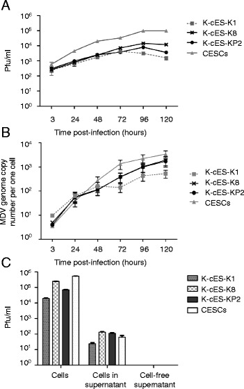
Fig. 2.

MDV replication in K-cESCs. Kinetics of infection of the vBAC20UL17mRFP on K-cESCs and CESCs, determined every 24 h from 3 to 120 h. Titres determined from two wells are given either in pfu/ml (a) or in MDV genome copy number/cell by qRT-PCR (b). Bars represent the mean ± SEM of viral loads. c Infectivity of the vBAC20UL17mRFP from K-cESCs or CESCs cells floating in the supernatant and from cell free supernatants. Statistical analyses were performed by Kruskal-Wallis one-way ANOVA test followed by two-tailed Mann–Whitney test. Analyses were done by using the software GraphPad Prism 5. No statistical significant difference between K-cESCs and CESCs was observed
