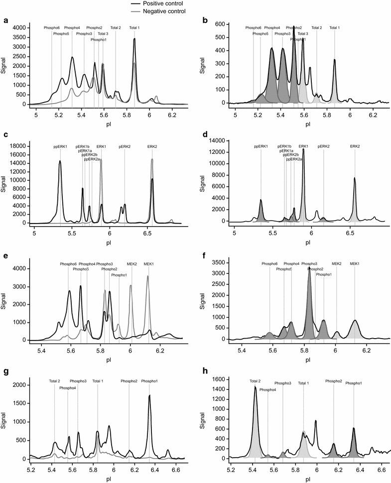
Fig. 3.

Representative NanoPro1000 chemiluminescence spectra for run controls and patient samples. Spectra overlay of positive (blue) and negative (green) run controls for AKT, ERK1/2, MEK1/2, and c-MET (a, c, e, and g) are shown in order to visualize regulation of phosphorylation. Representative spectra of patient samples (b, d, f, and h) are shown, including area under the curve (AUC) for unphosphorylated isoforms (light gray) and phosphorylated isoforms (dark gray)
