
Figure 1.
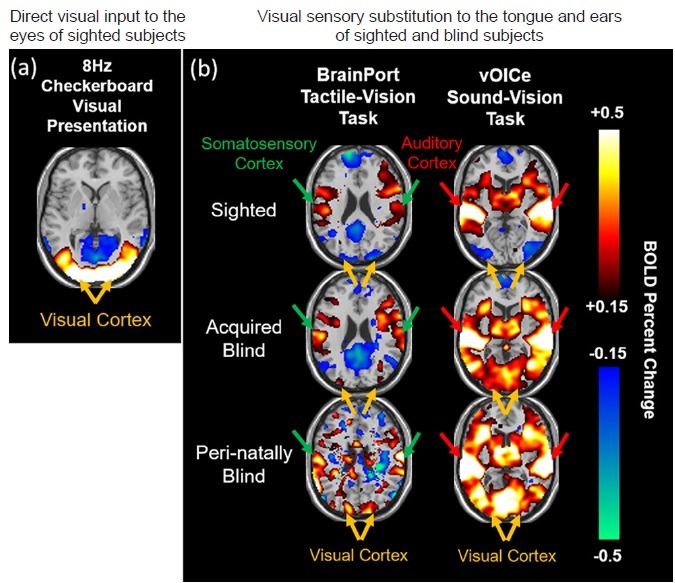
BOLD functional MRI of multimodal sensory substitution.
Task-based BOLD functional MRI (BOLD-fMRI) of the brain during (a) direct visual input to the eyes of sighted subjects via 8 Hz checkerboard visual presentation, and (b) visual sensory substitution to the tongue (left column) and ears (right column) of the same sighted and blind subjects via BrainPort tactile-vision (left column) and The vOICe sound-vision tasks (right column). BOLD percent change represents the amount of brain BOLD signal changes during task periods relative to resting periods. Note the positive BOLD responses in the visual cortex of sighted subjects in (a), and in the somatosensory cortex (left column) and auditory cortex (right column) of both sighted and blind subjects in (b) reflective of the subjects’ perception of vision, touch and auditory sensations respectively. In (b), the visual cortex of sighted subjects showed negative BOLD responses upon both tactile and auditory sensory substitution tasks, suggestive of cross-modal deactivations or inhibitions, whereas the visual cortex of blind subjects showed positive BOLD responses to both tactile and auditory vision substitution tasks similar to sighted subjects’ brain reactions to visual presentations in (a), indicative of the flexibility of the visual cortex to multi-sensory cross-modal plasticity upon visual deprivation. BOLD: Blood-oxygenation-level-dependent; MRI: magnetic resonance imaging.