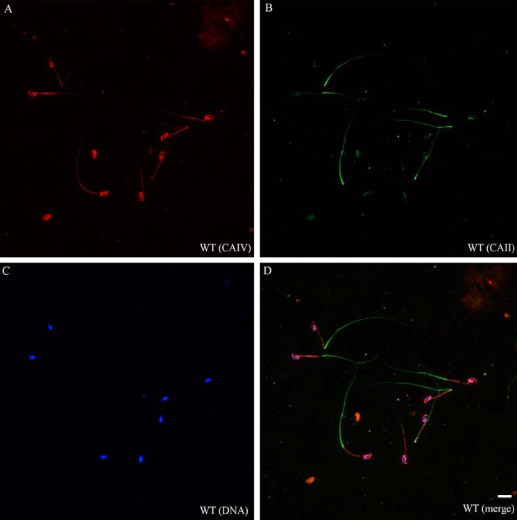
FIGURE 3.
CAII and CAIV have distinct localizations in murine spermatozoa. A–D, double immunofluorescence on fixed WT sperm indicates a CAII signal (green) in the principal piece of the sperm tail, whereas CAIV (red) is present in the acrosome and the plasma membrane of the entire sperm tail, predominantly in the mid-piece. CAII−/− CAIV−/− sperm did not show any signal (data not shown). Nuclei were stained with DAPI. Scale bar = 10 μm.

