
FIGURE 6.
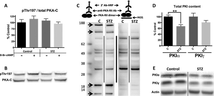
PKI is decreased in diabetic mice, but PKA phosphorylation and AKAP binding are unchanged. A and B, densitometry and Western blots of cardiac homogenates. The phosphorylated form of PKA standardized to total PKA. C, schematic and representative AKAP screen blot by PKA-RII overlay. PKA-RII bound to HRP via primary and secondary antibodies (left). Binding-deficient PKA-RII construct made by the addition of Ht31 (right) to show nonspecific bands. Arrows represent RII-AKAP interactions. D and E, PKI content shown by densitometry and representative Western blots. Standardized to actin. n = 5 for all data figures. **, p < 0.005, unpaired Student's t test.