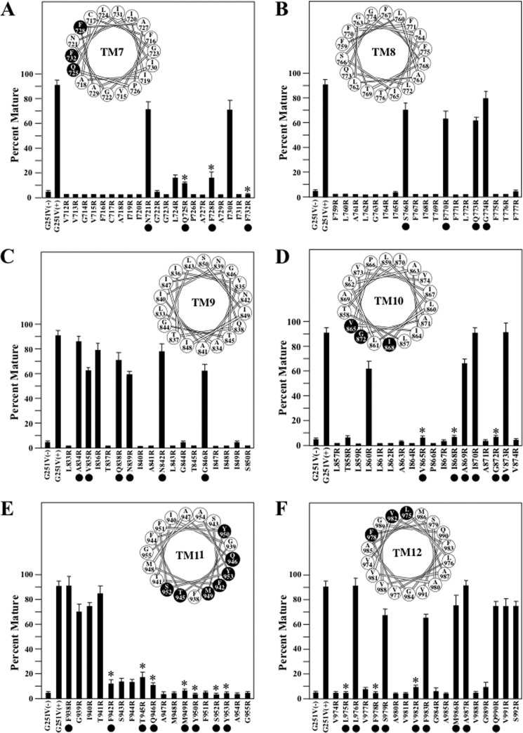
FIGURE 6.
Rescue of G251V arginine mutants in TMs 7–12 with tariquidar. Whole cell SDS extracts of cells expressing the G251V mutants containing arginines at various positions in predicted TM7 (A), TM8 (B), TM9 (C), TM10 (D), TM11 (E), or TM12 (F) in the presence of 0.5 μm tariquidar or the G251V parent in the absence (−) or presence (+) of tariquidar were subjected to immunoblot analysis. The amount of mature P-gp (170-kDa protein) relative to total (mature 170-kDa plus immature 150-kDa protein) (Percent Mature) was quantified. Each value is the mean ± S.D. (n = 3–5). The black dots identify residues predicted to line the drug-binding pocket shown in Fig. 5. An asterisk indicates a significant decrease (p < 0.001) relative to the G251V parent grown with tariquidar. The positions of the residues in the TM segments when arranged as α-helical wheels are shown in the insets. The black-filled circles represent arginine mutations predicted to line the drug-binding pocket and show significant difference relative to the G251V parent.

