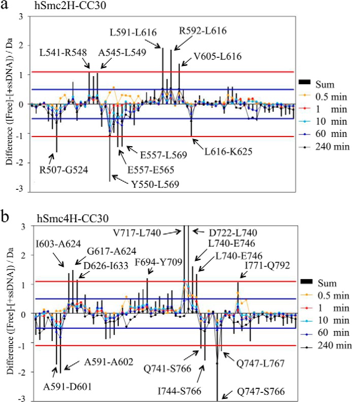
FIGURE 7.

Differential map of the hSmc2–4 hinge with middle coiled coils. For each peptide, the deuterium uptake at each exposure time of hSmc2–4 hinge with middle coiled coils in the presence of ssDNA (+ssDNA) was subtracted from that in the absence of ssDNA (Free). The summation of changes in the uptake over all exposure times has also been indicated. Positive or negative values indicate that the uptake rates were reduced or enhanced, respectively, upon interaction with ssDNA. Red lines, when the sum of difference is over 1.1 Da between two states for a peptide, the result reflects differing amide proton environments of the region covered by the peptide in solution, with 98% confidence. Blue lines, a difference of over 0.5 Da (at a specific exposure time) in a peptide indicates the differing amide proton environments of the region. a, hSmc2H-CC30 in the heterodimer. b, hSmc4H-CC30 in the heterodimer.
