FIGURE 6.
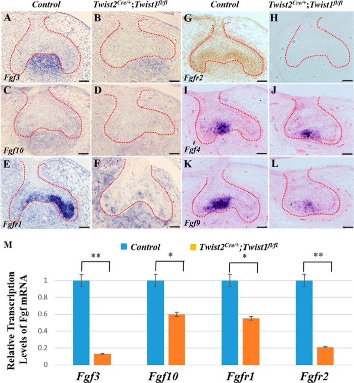
Reduced expression of Fgf and Fgfr in Twist2Cre/+;Twist1fl/fl mice. In situ hybridization was performed to determine the expression of Fgf3, Fgf10, Fgfr1, Fgf4, and Fgf9 in the developing control and Twist2Cre/+;Twist1fl/fl molars at E15.5 (A–L, signal in blue). Fgf3 and Fgf10 transcripts were observed in the dental mesenchyme, and Fgfr1, Fgf4, and Fgf9 transcripts were primarily found in the dental epithelium. The expression of Fgf3, Fgf10, Fgfr1, Fgf4, and Fgf9 was reduced dramatically in Twist2Cre/+;Twist1fl/fl molars compared with the control molars. Immunohistochemical staining showed that the Fgfr2 protein (signal in brown) was localized in both the dental epithelium and mesenchyme in the control molars (G) but was barely detectable in the Twist2Cre/+;Twist1fl/fl molars at E15.5 (H). The dashed lines in A–L indicate the boundary between the dental epithelium and mesenchyme. Quantitative PCR analysis also showed that the Fgf3, Fgf10, Fgfr1, and Fgfr2 transcripts were reduced remarkably in the Twist2Cre/+;Twist1fl/fl molars compared with the control molars at E14.5 (M). Error bars indicate mean ± S.E. *, p < 0.05; **, p < 0.01. Scale bars = 50 μm.

