
FIGURE 3.
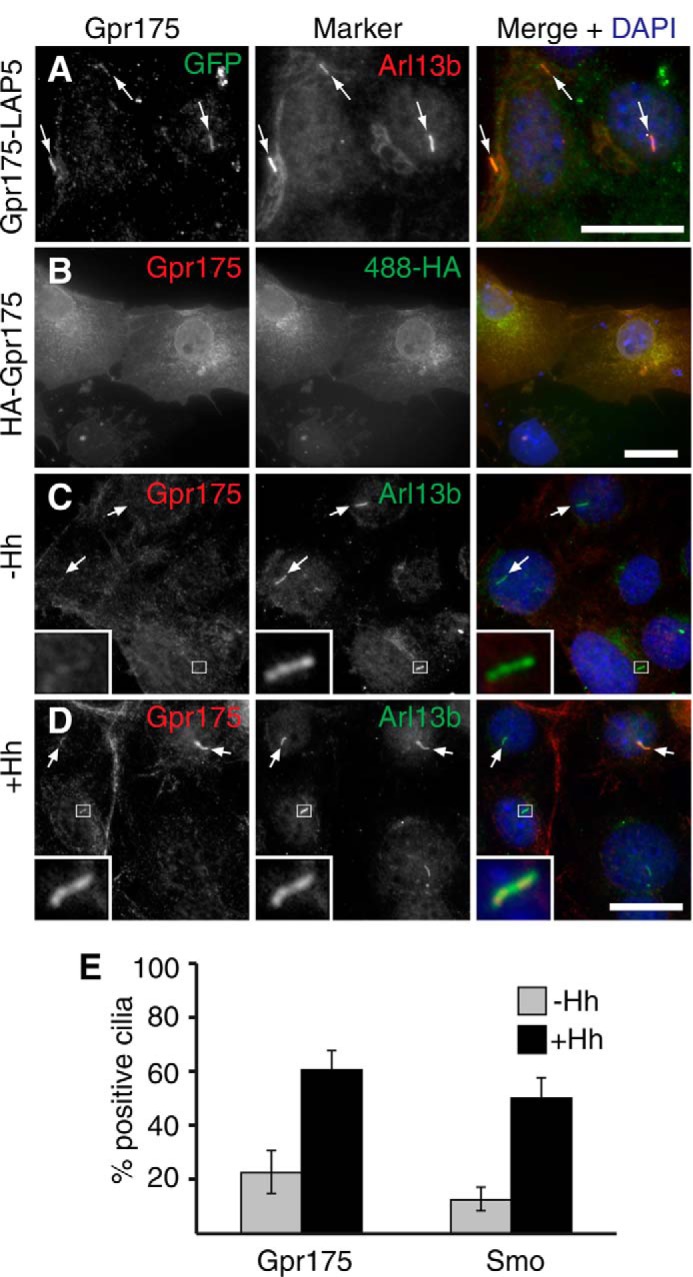
Gpr175 localizes to primary cilia of S12 cells in a Hh-dependent manner. A, NIH-3T3 cells stably expressing Gpr175-LAP5 were grown to confluence and serum starved for 16 h (without Hh) to induce ciliation. Cells were fixed and co-stained with mouse anti-GFP to detect Gpr175 (green, left) and rabbit anti-ARL13b (red, center) to label cilia. B, anti-human GPR175 monoclonal antibody 6H2 cross-reacts with HA-tagged murine Gpr175 transiently transfected in COS cells. 6H2 (red, left) colocalizes with anti-HA (green, center) at the plasma membrane and in the secretory pathway. Similar 6H2 staining was obtained in the absence of the anti-HA antibody, confirming it was not a cross-reactivity artifact (data not shown). C and D, S12 cells were fixed and stained with anti-GPR175 6H2 (red) and anti-ARL13b (green) following 24 h serum starvation alone (C) or with Hh stimulation (D). Scale bars are all 20 μm and the insets show ×5 magnification of the boxed area. The merged channels with nuclear counterstain DAPI (blue) are shown in the right column and arrows indicate cilia. Gamma levels were adjusted over the whole image to optimize the appropriate signals where necessary. E, the percentage of Arl13b-demarcated cilia positive for Gpr175 staining was scored, revealing that Gpr175 was found in only about 20% of cilia in the absence of Hh (gray), but in 60% following overnight Hh stimulation (black), very similar to the behavior of Smoothened (mean ± S.D. of 2 experiments, each counting ≥100 cilia, is shown).