
FIGURE 6.
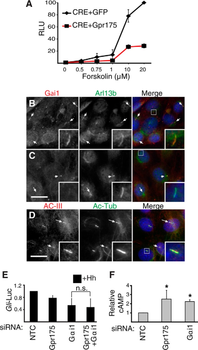
Gpr175 modulates cAMP levels via Gαi signaling. A, HEK-293T cells expressing CRE (cAMP response element)-luciferase along with a GFP (negative control, diamonds) or Gpr175 expression construct (squares, red line) were treated with varying doses of forskolin, thus increasing intracellular cAMP. The mean ± S.D. of four independent experiments normalized to the maximum tested (20 μm) Fsk in GFP-transfected cells are shown. RLU, relative luminescence units. B, S12 cells were serum starved for 24 h in the absence of Hh, fixed, and processed for immunofluorescence using mouse anti-Gαi1 antibody (red) and rabbit anti-ARL13b (green); merge with DAPI nuclear stain (blue) is shown in the right panel. C, as in B following siGαi1 transfection. D, S12 cells co-stained with rabbit anti-adenylyl cyclase-III(red), mouse anti-acetylated tubulin antibody (green), and DAPI (blue) in the merged right panel. Scale bars are 20 μm and insets show ×4 magnification of the boxed cilia. E, Gli-luciferase (Gli-Luc) levels in S12 cells following depletion of Gαi1 with or without siGpr175 in the presence of Hh (normalized to siNTC +Hh). Mean ± S.D. of 3 independent experiments are shown (n.s., not significant). F, S12 cells transfected with siNTC, siGpr175, or siGαi1 were serum starved for 24 h and then subjected to total cAMP measurement. Mean ± S.D. of three independent experiments are normalized to NTC control (*, p ≤ 0.05).