
FIGURE 8.
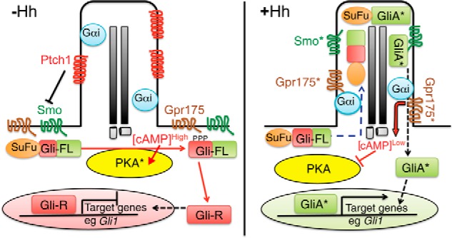
Working model for Gpr175 function as an enhancer of Hh signaling. Left panel, in the absence of Hh, Ptch1 in the primary cilium suppresses the activity of Smo and prevents its localization to cilia. Gpr175 is not present in cilia (its depiction on the plasma membrane is speculative) and so does not interact with ciliary Gαi, leading to high local concentrations of cAMP. cAMP stimulates PKA (asterisks denote active forms of all proteins) to phosphorylate full-length Gli3 (and Gli2; Gli-FL), which triggers proteasome-dependent cleavage of Gli3 into its repressor form (Gli-R). Gli3-R represses transcriptional activation of the Hh pathway target genes, thus the pathway is off. T-bars indicate inhibition; arrows indicate stimulation. Right panel, in the presence of Hh stimulation, Ptch1 is removed from cilia, allowing Smo to enter cilia and become activated. This leads to accumulation of Gpr175 in cilia, where it interacts with Gαi to inhibit local cAMP production, preventing PKA activity and Gli3 cleavage. The SuFu·Gli-FL complex accumulates at the tips of primary cilia, dissociates, and activated Gli-FL (GliA*) then exits cilia and enters the nucleus to promote transcription of Hh target genes such as Gli1. It is not clear if Gpr175 undergoes an activation step (like Smo) in addition to ciliary translocation to exert its activity.