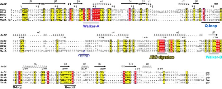
FIGURE 6.
Sequence alignment of SufC with various ABC ATPases from E. coli: HisP, BtuD, and MalK as ABC transporters and UvrA as a SMC protein. Red and yellow indicate identical and similar residues, respectively. Secondary structures of SufC are shown above the alignment with spirals (α-helices) and arrows (β-strands). Motifs conserved in ABC ATPases are shown below the alignment. Residues 737–792 in UvrA, an unrelated region, are omitted from the sequence.

