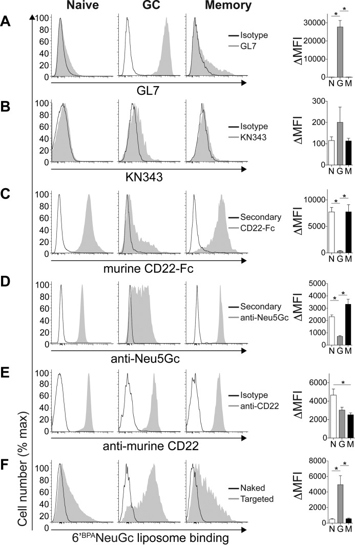
FIGURE 3.
Unmasking of CD22 on murine GC B-cells relative to naive and memory B-cells. Flow cytometry staining of naive (IgD+CD38+CD95−), GC (CD38−CD95+), and memory (IgD−CD38+CD73+CD80+) murine CD19+ B-cells with GL7 (A), KN343 (B), murine CD22-Fc (C), anti-NeuGc (D), anti-murine CD22 (E), and fluorescent liposomes (F) displaying no ligand (naked) or a selective murine CD22 ligand (6′BPANeu5Gc; targeted). Quantitated ΔMFI (mean fluorescence intensity) values represent background-subtracted values and represent the average and standard error of three replicates and is representative of three independent experiments. *, p < 0.05; N, naive; G, germinal center; M, memory.

