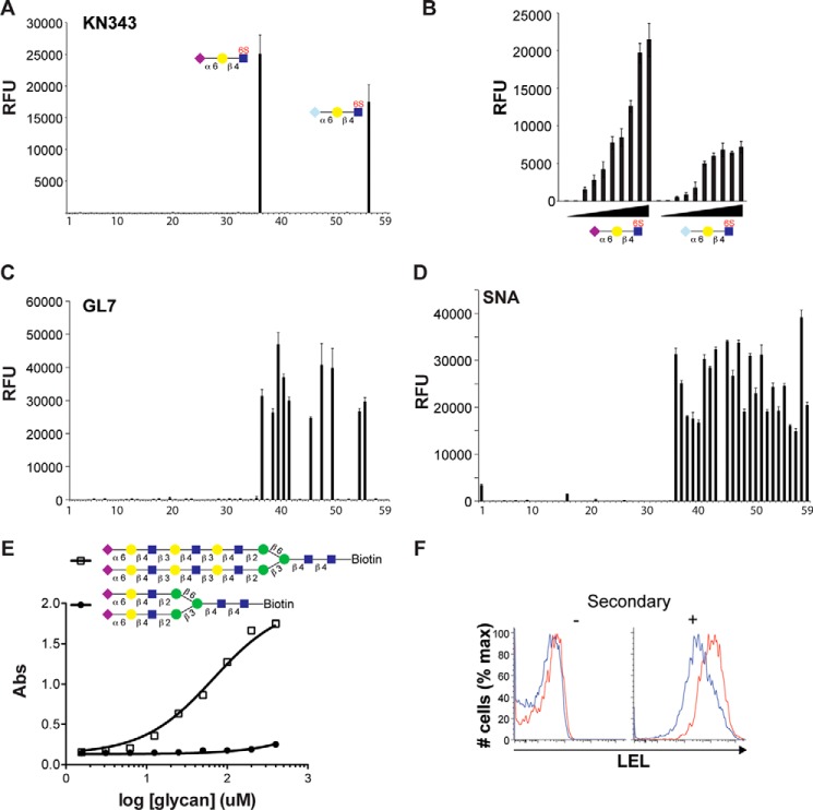
FIGURE 6.
Glycan binding specificity of the KN343 and GL7 antibodies. A, probing the 59 glycan sialoside microarray with KN343 demonstrates that it selectively recognizes Neu5Acα2–6Galβ1–4(6S)GlcNAc and Neu5Gcα2–6Galβ1–4(6S)GlcNAc. B, printing concentration of Neu5Acα2–6Galβ1–4(6S)GlcNAc and Neu5Gcα2–6Galβ1–4(6S)GlcNAc was titrated in 2-fold serial dilutions and probed with KN343. C and D, probing a 59 glycan sialoside microarray with GL7 (C) and SNA (D) demonstrates that GL7 selectively recognizes glycans containing terminal α2–6-linked Neu5Ac and prefers structures containing more than one LacNAc repeat. A complete list of glycans is described in Fig. 5. E, direct binding of a complex N-glycan containing 1 (closed circles) or 3 (open squares) LacNAc units per arm to GL7 by ELISA. F, staining of naive (CD19+CD95−CD38+GL7−; blue) and GC (CD19+CD95+CD38−GL7+; red) B-cells for LEL. The histogram on the left represents background staining without secondary antibody. RFU, relative fluorescence units.

