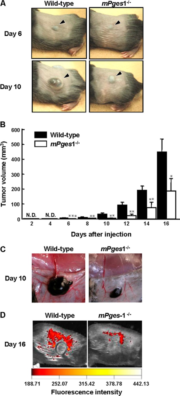
FIGURE 1.

Tumor growth was attenuated in mPges1−/− mice injected with B16 cells. A, B16 cells were injected into the dorsal subcutaneous tissue of wild-type and mPges1−/− mice. Representative pictures of the tumors on days 6 and 10 after injection are shown. The arrowheads indicate the subcutaneous solid tumor. B, the tumor volume was determined using calipers. *, p < 0.05; **, p < 0.01; ***, p < 0.001. N.D., not detected. Data are mean ± S.E. of wild-type (n = 9) or mPges1−/− (n = 6) mice. C, representative pictures of new blood vessels around dorsal subcutaneous tumors in mPges1−/− or wild-type mice on day 10 after injection. D, fluorescence imaging of new blood vessels around dorsal subcutaneous tumors in mPges1−/− or wild-type mice on day 16 after injection.
