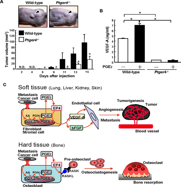
FIGURE 7.
Tumor growth and VEGF production are attenuated in Ptger4−/− mice. A, Ptger4−/− or wild-type mice were injected with B16 cells into the dorsal subcutaneous tissue. Representative pictures of the subcutaneous tumors on day 9 after injection are shown in the top panel. The arrowheads indicate a subcutaneous solid tumor. The tumor volume was determined, and the data are shown in the bottom panel as mean ± S.E. of the wild-type (n = 9) or Ptger4−/− (n = 8) mice. *, p < 0.05. N.D., not detected. B, dermal fibroblasts from Ptger4−/− or wild-type mice were cultured in the presence or absence of PGE2 (10 μm). After 48 h, the concentration of VEGF-A in the conditioned media was determined by ELISA. Data are mean ± S.E. of three wells. *, p < 0.001. C, schematic of the PGE2/EP4-depedendent mechanism involved in angiogenesis, tumorigenesis, and osteoclastogenesis. In soft tissues, stromal fibroblasts interact with cancer cells and produce PGE2 via mPGES-1, and then PGE2 induces VEGF-A and bFGF production as a result of autocrine/paracrine signaling via EP4. In hard-tissue bone, the interaction with cancer cells elicits mPGES-1-dependent PGE2 production by osteoblasts, and then PGE2 induces the expression of RANKL on the surface of osteoblasts to induce osteoclast formation, which is critical for the osteolysis associated with the bone metastasis of cancer. AA, arachidonic acid.

