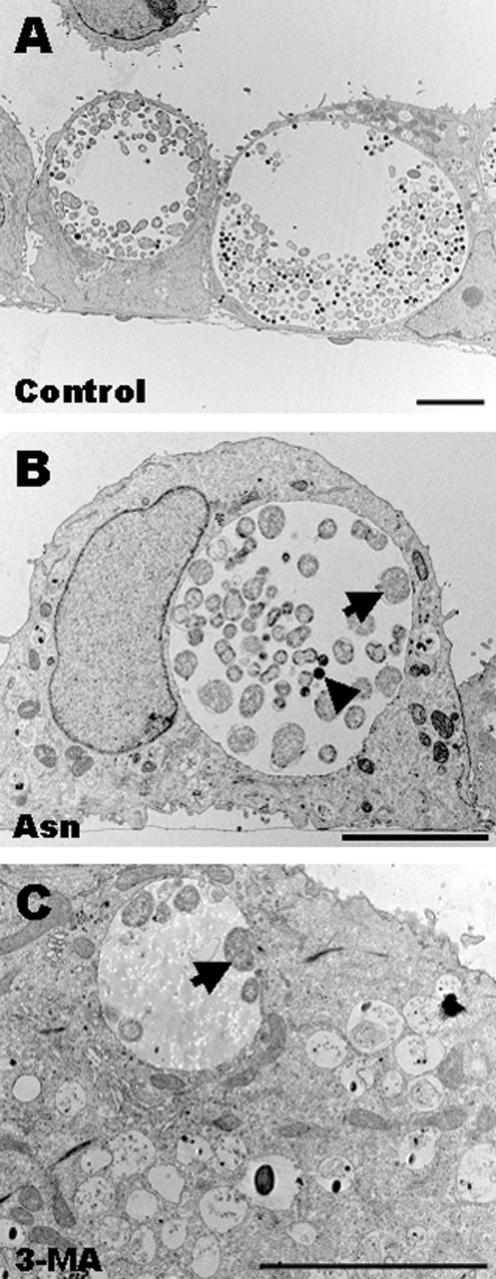
FIG. 3.
C. trachomatis L2 inclusions in cell monolayers exposed to 3-MA or exogenous excess Asn, as viewed by EM. Host cells were pretreated with one of the chemicals for 30 min and then infected for 32 h in the presence of the respective chemical. (A) Control inclusions developed for 32 h in medium without additives contained normal forms of chlamydiae. (B) Inclusions developed in the presence of 30 mM Asn. Inclusions here are less mature and contain mostly RBs (arrow) compared to control inclusions (see panel A). Few EBs were observed (arrowhead). (C) Inclusions grown in the presence of 5 mM 3-MA contained low number of chlamydial forms, which were exclusively RBs (arrow). Bars, 5 μm.

