Figure 3.
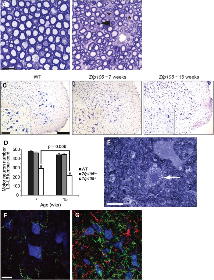
Motor neuron and axonal degeneration and gliosis in Zfp106−/− mouse spinal cord. (A and B) Representative toluidine blue stained sections of lumbar L4 and L5 ventral roots from 15-week old WT (A) and Zfp106−/− (B) mice. Zfp106−/− mice have fewer motor axons than WT littermates (spaces are marked with asterisks), and have degenerating axons, indicated with arrow head. Scale bar is 20 μm. (C) Representative images of the ventral horn of lumbar spinal cord sections stained for Nissl from WT, 7- and 15-week-old Zfp106−/− mice. The number of motor neurons in the sciatic pool (inset) was counted for each genotype and age to calculate motor neuron survival (D). Scale bars: main images 200 μm inset 100 μm. (D) The survival of motor neurons in the sciatic pool in female mice at 7- and 15-weeks of age. Motor neuron survival in 7-week-old Zfp106−/− mice (294 ± 11) is reduced compared with WT littermates (481 ± 9) and further reduced in 15-week-old Zfp106−/− mice (217 ± 9). Numbers represent the mean ± SEM. At least five animals were assessed per genotype per time point (*P < 0.001). (E) Representative toluidine blue stained section of 15-week-old Zfp106−/− lumbar spinal cord showing an example of a vacuolated (arrow) motor neuron. (F and G) Lumbar spinal cord sections of 15-week-old WT (F) and Zfp106−/− (G) mice stained for IBA-1 (green), GFAP (red) and Nissl (blue). Immunoreactivity for micro- and astrogliosis is dramatically increased in the lumbar of 15-week-old Zfp106−/− mice compared with WT littermates. Scale bar is 20 μm.

