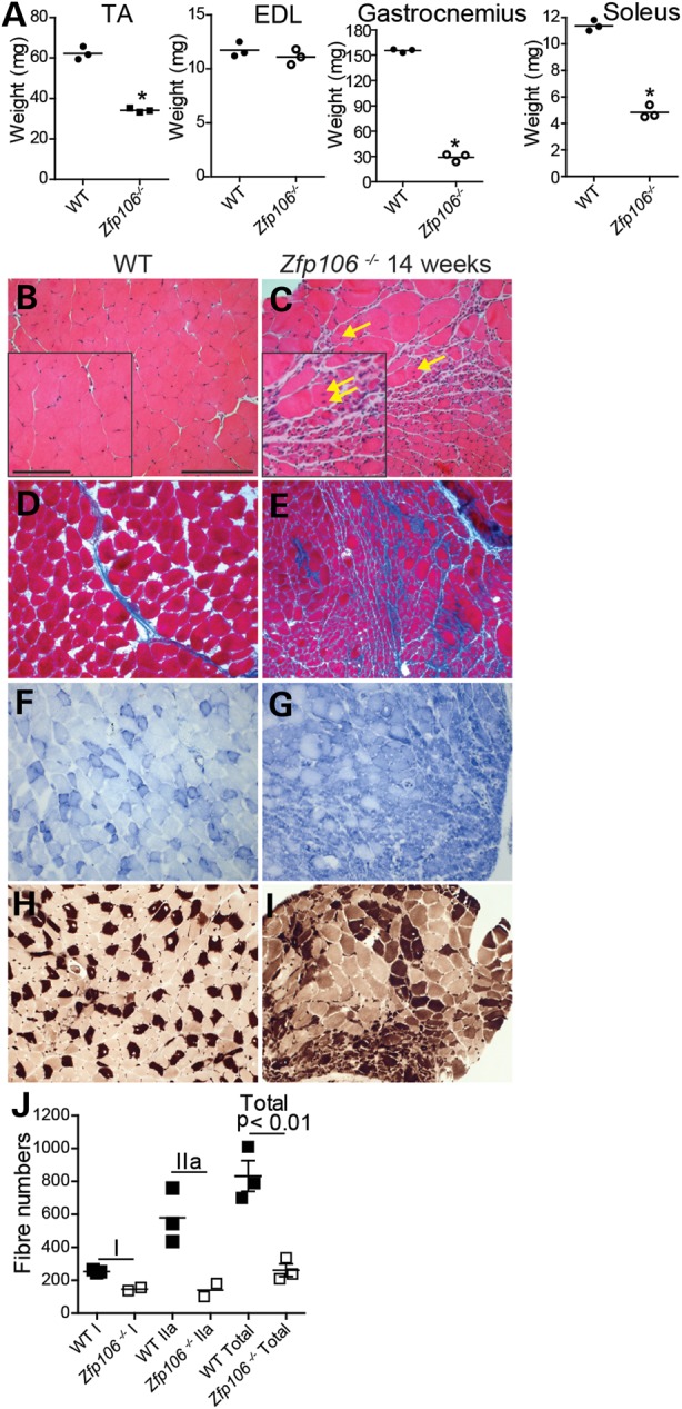
Figure 6.

Muscle histology of WT and Zfp106−/− male mice. (A) Muscle weights for the TA (WT, 62 mg ± 1 mg; Zfp106−/−, 34 mg ± 1 mg), EDL (WT, 11 mg ± 0.4 mg; Zfp106−/−, 11 mg ± 0.4 mg), gastrocnemius (WT, 155 mg ± 1 mg; Zfp106−/−, 29 mg ± 23 mg) and soleus (WT, 11 mg ± 0.2 mg; Zfp106−/−, 5 mg ± 0.3 mg) from male 14-week old WT and Zfp106−/− littermates (n = 3). TA, gastrocnemius and soleus weights are significantly different from WTs (*P < 0.01). (B–I) Representative images of muscle histopathology from 14-week-old WT and Zfp106−/− mice: (B and C) H&E staining of the gastrocnemius shows significant atrophy, reduced fibre size and centralized nuclei (yellow arrows) in Zfp106−/− muscle compared with WT littermates; (D and E) Masson's trichome staining of gastrocnemius muscle reveals fibrosis in Zfp106−/− muscle (E), evident with increased blue staining; (F and G) NADH-TR staining of the gastrocnemius shows greatly increased dark blue staining in Zfp106−/− muscle (G) compared with WT littermates, indicative of a higher proportion of slower twitch, type I muscle fibres; (H and I) ATPase staining of Zfp106−/− soleus muscle (I) showing an increase in darkly stained fibres, indicating an increase in the proportion of type I fibres overall. Scale bars: main images 20 μm, inset 10 μm. (J) Type I (WT, 253 ± 6 fibres; Zfp106−/−, 147 ± 10 fibres), type IIa (WT, 579 ± 95 fibres; Zfp106−/−, 141 ± 40 fibres) and total fibre number (WT, 833 ± 92 fibres; Zfp106−/−, 261 ± 38 fibres) were counted for the soleus muscle from 14-week-old WT and Zfp106−/− littermates using ATPase staining. The soleus from Zfp106−/− mice has significantly fewer muscle fibres than that of WT mice (P < 0.01).
