
Figure 2.
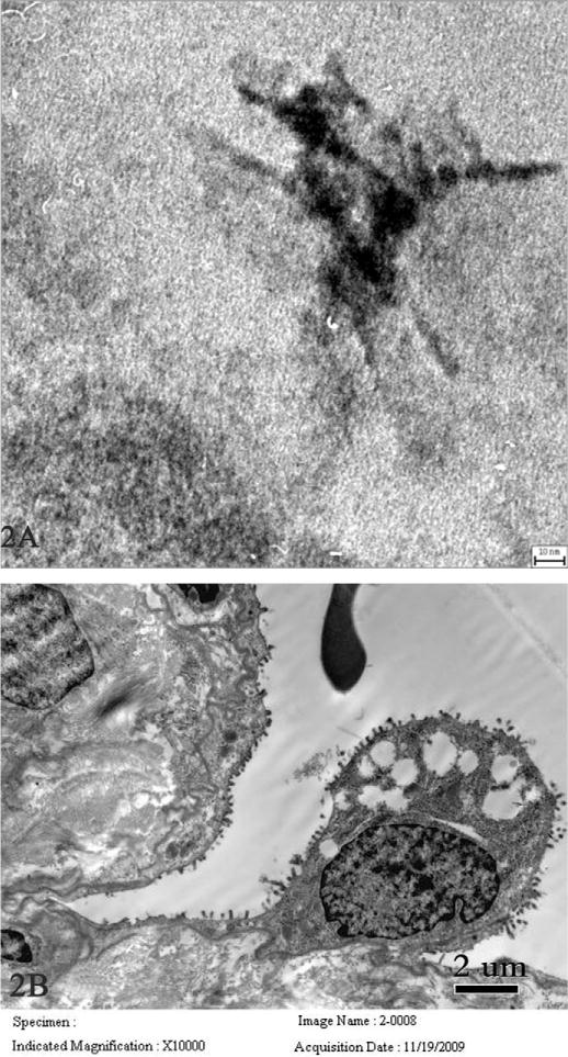
Transmission electron microscopy images of fibrous nanostructured bodies and pulmonary damage in another nineteen-year-old female worker who died eighteen months after symptom onset. Fibrous nanostructured bodies ~70 nm in length were observed in the nuclei of an alveolar epithelial cell, where the nanostructure is in tangled form (A, scale bar = 30 nm). B (scale bar = 2 μm) shows a fraction of a red blood cell in alveolar spaces, hyperplasia, and vacuolar degeneration of type II pneumocytes with thinning and loss of surface microvilli.