
Figure 4.
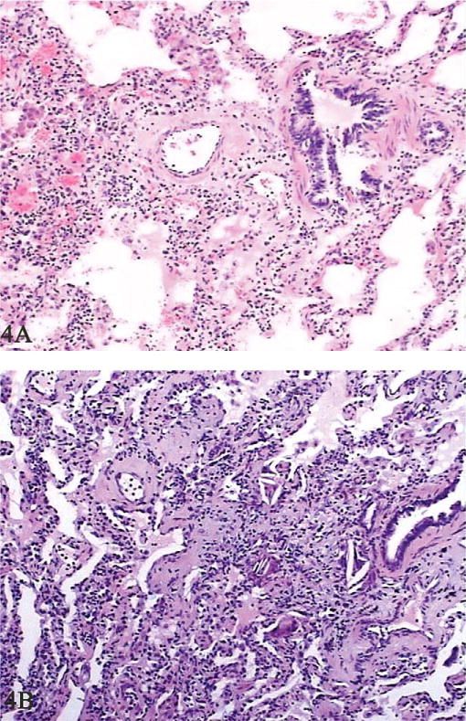
Histological changes in patients’ lungs. In the early stage, aggregation of macrophages in the alveolar space, swollen and widened alveolar septa, and pulmonary fibrosis were observed (A, hematoxylin and eosin stain, 100×). In the later disease process, pulmonary alveoli were partly emphysematous with aggregations of macrophages and proliferations of type II alveolar epithelial cells, and the alveolar septum was widened with blood vessel dilatation and congestion (B, hematoxylin and eosin, 100×).