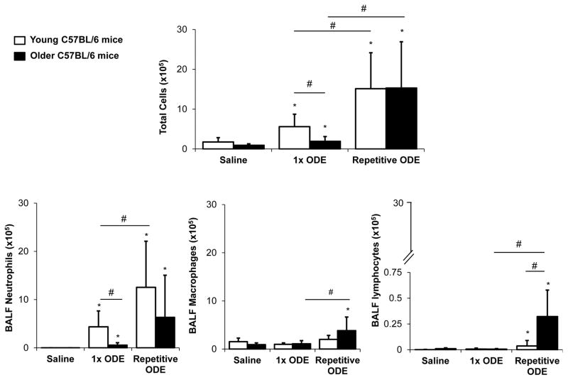
Figure 1. Airway inflammatory cellular influx following organic dust extract (ODE) exposure in young and older mice.
Young (7–9 week) and older (12–14 month) C57BL/6 mice were treated with ODE once (1x) or daily for 3 weeks (repetitive) using i.n. inhalation method. Five hours following final exposure, mice were euthanized and bronchoalveolar lavage fluid (BALF) was collected. Mean with standard deviation bars of total cells, neutrophils, macrophages, and lymphocytes from each treatment group is shown (n=9–12 mice/group from a minimum of 2 independent studies). Statistically significant (*p<0.05) vs. saline. Lines between groups denote statistical significance (#p<0.05).

