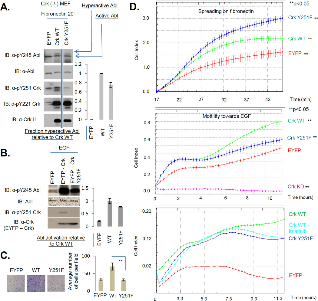
Figure 5. Phosphorylation of the Crk SH3C at Y251 inhibits cell spreading on fibronectin and promotes cell motility towards EGF.
A, Crk (−/−) MEFs stably reconstituted with EYFP, Crk WT or Crk Y251F were plated on fibronectin coated dishes for 20 minutes. Lysates were analyzed by western blotting with the antibodies shown. Quantification of pY245 on hyperactive Abl is shown on the right. All samples were run on the same gel and hence are comparable. However, the EYFP and WT lanes were not adjacent to the Y251F lane and hence have been demarcated by straight lines at the edges. B, MDA-MB-468 cells stably expressing EYFP, EYFP-Crk WT, EYFP-Crk Y251F were stimulated with EGF for 5 minutes and lysates were analyzed by western blotting with the antibodies shown. Quantification of pY245 Abl normalized to total Abl is shown on the right. C, Cells in B were analyzed for their motility towards EGF in a transwell Boyden chamber assay. ** indicates p<0.05. Representative images are shown on the left. D, Top, Cells in A were plated on fibronectin coated E-plates in triplicate and real-time cell spreading was recorded using xCelligence. Shown is a representative of two independent experiments each performed in triplicate. Quantification of cell index is shown as mean +/− SD. ‘**’ indicates p<0.05. Middle and Bottom, Cells in B in addition to MDA-MB-468 stably expressing Crk shRNA (Crk KD) were seeded into CIM-plates in quadruplicate in the middle panel and in duplicate in the bottom panel with an EGF gradient of 10 ng/ml and cell motility was recorded in real time using xCelligence. Middle panel is a representative of four independent experiments each performed in quadruplicate while the bottom panel is a representative of two independent experiments each performed in duplicate. Quantification of cell index is shown as mean +/− SD in the middle panel and as average of duplicates in the bottom panel. ’**’ indicates p<0.05.

