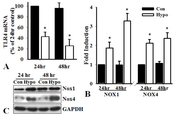
Figure 5.
Hypoxia decreases the expression of TLR4 and induces Nox1 and Nox4. PASMC from WT mice were seeded at 80% confluence and exposed to control (20% oxygen, Con) or hypoxia (2% oxygen, Hypo) condition for 24 and 48 hours. Real-time PCR analysis of the expression of A). TLR4 using primers for mouse TLR4: F-5′-GCTTTCACCTCTGCCTTCAC; R-5′-CGAGGCTTTTCCATCCAATA; B). Nox1 and Nox4 using specific primers shown in Fig 4. Results shown are the expressions of TLR4, Nox1 and Nox4 mRNA (normalized by GAPDH mRNA) as percentage of that in cells exposed to condition for 24 hours, defined as 100% (n=3, *p<0.05). C). Western blot analysis of the expression of Nox1 and Nox4 proteins. The expression of GAPDH was used as a loading control.

