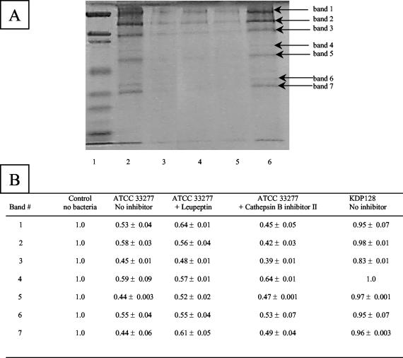
FIG. 5.
Degradation of Matrigel constituents by cells of P. gingivalis ATCC 33277 and KDP128, as determined by SDS-PAGE analysis and Coomassie blue staining. (A) Lanes: 1, molecular weight markers; 2, Matrigel alone; 3, Matrigel plus ATCC 33277; 4, Matrigel plus ATCC 33277 and leupeptin; 5, Matrigel plus ATCC 33277 and cathepsin B inhibitor II; 6, Matrigel plus KDP128 (rgpA rgpB kgp). (B) The relative intensities of bands were assessed by using Scion Imaging Software. A value of 1 was given to each band of the Matrigel control. Results are reported as means and SDs.

