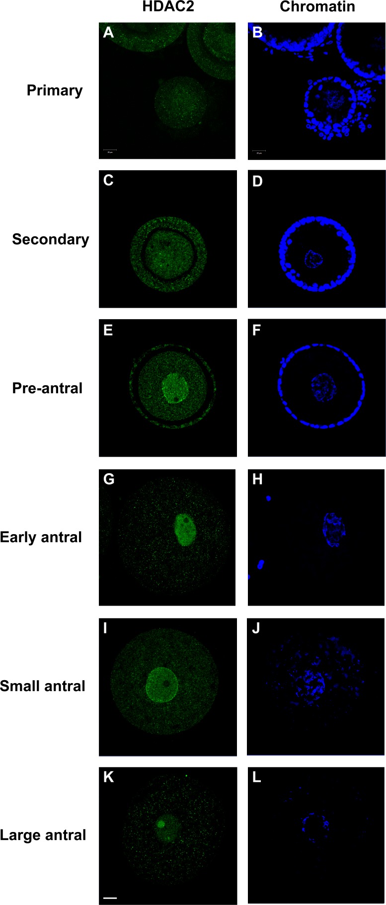
FIG. 2.
Expression patterns of HDAC2 in the cat GV within oocytes from different stages of intraovarian follicles. Confocal images of immunostaining of HDAC2 (A, C, E, G, I, and K) from primary stage to large antral stage, with the respective chromatin counterstained with Hoechst (B, D, F, H, J, and L). Oocytes from early, small, and large antral follicles were stripped of surrounding cumulus cells before evaluation. Bar = 20 μm.

