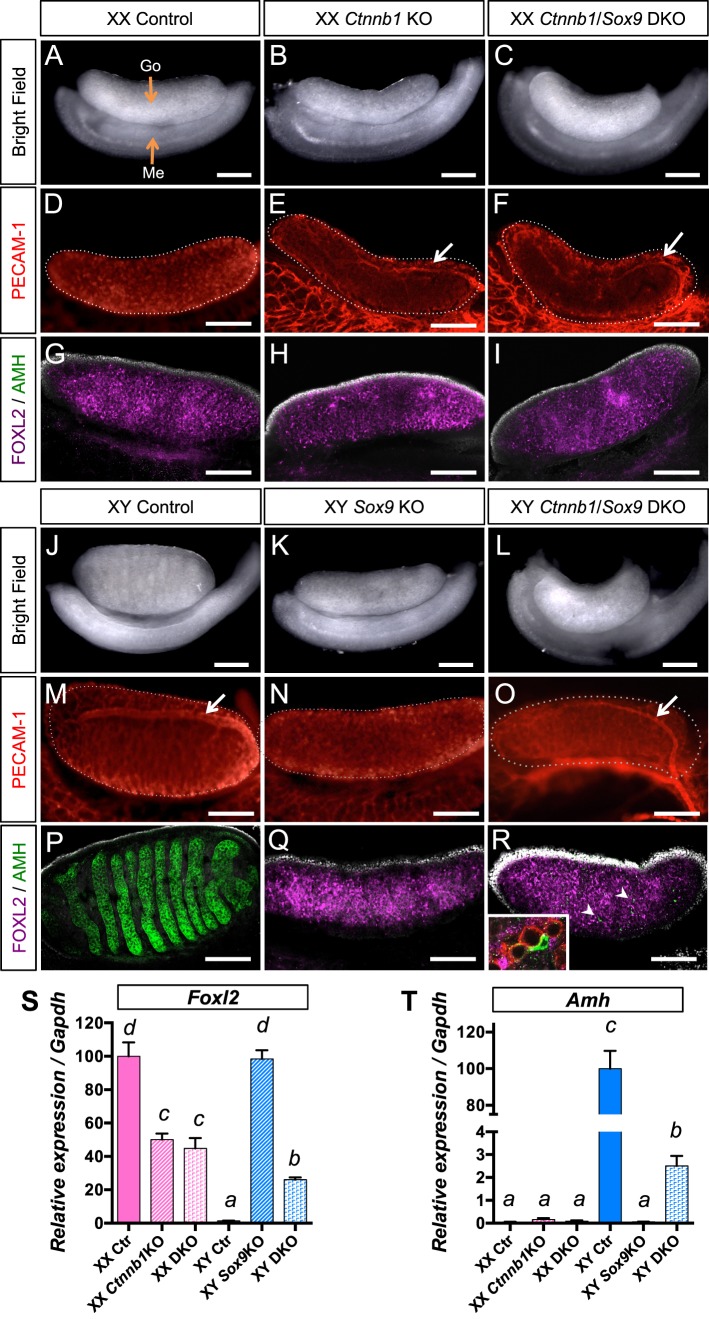
FIG. 1.
Consequences of inactivation of both β-catenin and Sox9 in the gonads at 14.5 dpc. The 14.5 dpc XX (A–I) and XY (J–R) gonads of various genotypes (control, Ctnnb1 KO, Sox9 KO, and Ctnnb1/Sox9 DKO) were analyzed by bright field microscopy (A–C, J–L), immunofluorescence for germ cell/vasculature marker PECAM-1 (D–F, M–O) and supporting cell markers FOXL2/AMH (G–I, P–R). White arrows show the testis-specific vessel at the surface of the gonad (E, F, M, O). Nuclei are labeled with 4′,6-diamidino-2-phenylindole ([DAPI] grey) in G–I and P–R. R) Arrowheads show the AMH+ cells, and the inset is a higher magnification of a part of the XY Ctnnb1/Sox9 DKO gonads that contain PECAM+ germ cells (red) surrounded by FOXL2+ granulosa cells (magenta) and one AMH+ Sertoli-like cell (green). Go, gonad; Me, mesonephros. The white dotted lines outline the gonads. Bars = 200 μm. S and T) represent quantitative PCR analysis of Foxl2 (S) and Amh (T) mRNA expression in 14.5 dpc gonads. Error bars represent SEM of five biological replicates (Student t-test, P < 0.05; a < b < c < d).

